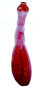
Echinococcus granulosus <(= Echinococcus unilocularis) is a parasitic tapeworm that has dogs and other canids (e.g. foxes, wolves, coyotes, etc.) as final hosts. It is also called the hydatid worm and the dog tapeworm (not to be confused with Dipylidium caninum).
But it infects many other domestic and wild mammals (mainly ungulates, but also rodents) that act as intermediate hosts, including cattle, sheep, goats, pigs, horses, dromedaries, South American camelids, deer, kangaroos, etc. where it causes a disease called cystic echinococcosis or hydatid disease. Humans can also act as intermediate hosts. There are a few reports that cats can acts as intermediate hosts as well.

Echinococcus granulosus is found worldwide, but the prevalence varies a lot. It is generally more abundant in rural regions with abundant livestock and wildlife together with poor sanitary conditions.
Echinococcus multilocularis is a related species that affects dogs, cats, and humans but not livestock.
Are dogs, horses or livestock infected with Echinococcus granulosus contagious for humans?
- YES. The worm eggs shed with the feces of dogs can contaminate food and drinking water and are infective for humans as well as other animals. Contact with contaminated dogs can also be contagious, because their hair coat can carry such eggs as well. See the life cycle below. Intermediate hosts (horses, livestock, etc.) contaminated with Echinococcus granulosus, i.e. suffering of cystic echinococcosis (= hydatid disease) are not contagious for humans.
You can find additional information in this site on the general biology of parasitic worms and/or tapeworms.
Final location of Echinococcus granulosus
Predilection site of adult Echinococcus granulosus worms is the small intestine.
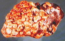
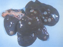
The hydatid cysts on intermediate hosts (cattle, sheep, pigs, etc.) develop mainly in the liver and the lungs but can affect other organs, e.g. the brain.
Anatomy of Echinococcus granulosus
Adult Echinococcus granulosus worms are rather small, not longer than 7 mm. They have only 4 segments, the last one being the largest and gravid, i.e. filled with eggs. The head (scolex) has 4 suckers and numerous hooks for attaching to the gut's wall. Otherwise, as other tapeworms, Echinococcus granulosus has neither a digestive tube, nor circulatory or respiratory systems. It doesn't need them because each proglottid absorbs what it needs directly through its tegument.
Each proglottid has its own reproductive organs of both sexes (i.e. they are hermaphroditic) and excretory cells known as flame cells (protonephridia). The reproductive organs in each proglottid have a common opening called the genital pore. In young proglottids all these organs are still rudimentary. They develop progressively, which increases the size of the proglottid as it moves towards the tail. Mature gravid proglottids are full of eggs and detach from the strobila (the chain of segments) to be shed outside the host with its feces.
The eggs are have an ovoid to spherical shape, are rather small (~30 micrometers). They are embryonated (i.e. contain already developed larvae called oncospheres or hexacanths) with a thick capsule radially striated.
The hydatid cysts (also known as metacestodes or bladder worms) develop in various organs of the intermediate hosts, are oval to spherical in shape and grow slowly but steadily. Eight weeks after infection the cysts can reach about 2.5 mm in diameter, three months later about 2 cm. Hydatid cysts found in slaughterhouses can reach the size of an orange (5 to 10 cm). Infected organs can have dozens of cysts. Each cyst is filled with liquid and contains several heads of the parasite (protoscolices) produced through asexual multiplication.
Life cycle and biology of Echinococcus granulosus
Echinococcus granulosus has an indirect life cycle with dogs (and other canids) as final hosts, and numerous domestic and wild mammals, including humans as intermediate hosts.
Gravid segments filled with eggs are shed with the dog's feces hosts. Each adult worm deposits one gravid segment aprox. every two weeks, and each gravid segment contains several hundred eggs. Once in the environment, survival of the eggs depends strongly on the climatic conditions and is lower by warm and dry weather. Infectivity decreases with time.

Intermediate hosts ingest such eggs through contaminated food (both fresh herbage and hey) or water. Inside the intermediate host, the eggs release the hexacanths (larvae) in the intestine. They go through the intestinal wall, get into the portal vein and are transported to the liver. The capillaries in the liver act like a filter that retains numerous hexacanths, which develop to cysticercoids and produce the hydatid cysts.
The blood stream can transport hexacanths to other organs too, e.g. to the lungs or the brain, where the respective capillary networks retain the hexacanths that produce the cysts. Such a cyst can rupture and the immature worms are transported further to other organs. The hydatid cysts in intermediate hosts can contain hundreds if not thousands of infective larvae.
The immature worms in the hydatid cysts do not complete development to adults there. They do it only once they get into a final host that has fed on a prey infected with hydatid cysts. In this case the young worms (protoscolices) are released after digestion of the cysts, attach to the gut's wall, complete their development to mature adults and start producing eggs.>
The prepatent period (time between infection and recovery of first eggs in the feces) is 6 to 7 weeks.
loadposition Native-Eng-2}
Harm caused by Echinococcus granulosus infections, symptoms and diagnosis
For dogs and other final hosts Echinococcus granulosus infections are benign, mostly without clinical signs, unless in case of very heavy infections, which are unusual.
<For livestock and other intermediate hosts such as horses, the development of hydatid cysts is mostly asymptomatic, unless the organs are heavily affected, which is usually due to the growing cysts pressuring the organ tissues. Parts of the tissue die, which impairs the functioning of the affected organ. The clinical signs depend on the affected organs. Digestive disturbances, cough and difficult breathing (dispnea) have been described. The major damage for livestock is organ condemnation at slaughter.
For humans as intermediate hosts, hydatid cysts can be a serious disease. The problem is that they develop very slowly over years and without symptoms. They are often discovered too late. If vital organs (e.g. brain, heart) are affected harm is often irreparable and fatalities are possible. Rupture of the cysts can also cause strong anaphylactic reactions.
Diagnosis in dogs cannot be based on egg identification in the feces, because Echinococcus eggs are undistinguishable from Taenia spp eggs. ELISA tests based on egg antigens in the feces are very reliable but not available in some countries. Newer PCR (Polymerase Chain Reaction) tests based on DNA from egg in the feces have also been developed.
Diagnosis in livestock or other intermediate hosts is usually made after slaughter or necropsy. So far there are no reliable biochemical tests for early detection.
Prevention and control of Echinococcus granulosus
In dogs
In endemic regions it is advisable to reduce the number of stray dogs. Domestic and working dogs must be kept away from contaminated offal. They can be preventatively treated with approved taenicides. These products contain mainly broad-spectrum anthelmintic active ingredients such as benzimidazoles (e.g. fenbendazole, febantel, mebendazole) or specific taenicides such as praziquantel and epsiprantel, the latter often in combination with nematicides (e.g.levamisole, milbemycin oxime, pyrantel, etc.) to cover a broader spectrum of worms. These preventative measures are highly recommended also for urban dogs in endemic areas, in order to prevent transmission of echinococcosis to humans.
Most of these pet wormers are available in formulations for oral delivery either as solids (tablets, pills, etc.) or as liquids (drenches, suspensions, etc.). There are also a few injectable and spot-on (= squeeze-on = pipettes) taenicides in some countries (mainly with praziquantel).
Most wormers kill the worms shortly after treatment and are metabolized and/or excreted within a few hours or days. This means that they have a short residual effect, or no residual effect at all. As a consequence treated animals are cured from worms but do not remain protected against new infections. To ensure that they remain worm-free the pets have to be dewormed periodically, depending on age and the local epidemiological, ecological and climatic conditions.
Other classic livestock anthelmintics such as macrocyclic lactones (e.g. ivermectin, selamectin, etc.), levamisole, tetrahydropyrimidines (e.g. pyrantel, morantel) and piperazine derivatives are not effective at all against Echinococcus species or whatever tapeworms.
So far there are no antiparasitic medicines for style="text-decoration: underline;">external use such as shampoos, soaps, sprays, powders, insecticide-impregnated collars, etc. that control established tapeworm infections.
In livestock and horses
Best prevention of Echinococcus infections in livestock and horse farms is to keep dogs away from feeding contaminated organs, especially working dogs. To achieve it is essential to thoroughly cook whatever offal they get, or to feed them on commercial dog food. Wild rodents (rats, mice, squirrels, etc.) can also be intermediate hosts of Echinococcus granulosus and dogs can become infected if they eat such infected preys. If this is not possible, working dogs should be regularly treated with taenicides as previously mentioned

A true vaccine against livestock echinococcosis caused by Echinococcus granulosus is now available in several countries. The commercial brand is called PROVIDEAN HIDATIL EG 95 ® and was introduced in 2011 in Argentina by a company called TECNOVAX. It is approved for use on cattle, sheep, goats and South American camelids. It is based on the EG95 antigen obtained from Echinococcus granulosus eggs. One dose provides up to 82% protection, two doses up to 97% and three doses 100% protection. Hopefully this powerful weapon will soon become available in other endemic regions.
The use of anthelmintics against hydatid cysts is usually not indicated in livestock, neither therapeutically (i.e. to cure already established hydatid cysts) nor prophylactically (i.e. to prevent the development of hydatid cysts). Most active ingredients with efficacy against tapeworms such as broad-spectrum benzimidazoles (e.g. albendazole, fenbendazole, febantel, mebendazole) or specific taenicides such as praziquantel are ineffective against Echnicoccus granulosus hydatid cysts at the usual therapeutic dose and have to be administered at significantly higher doses and repeatedly to achieve only partial control. This is either too expensive, or not well tolerated by livestock, or both.
Other classic livestock anthelmintics such as macrocyclic lactones (e.g. ivermectin, doramectin, moxidectin, etc.), levamisole, tetrahydropyrimidines (e.g. pyrantel, morantel) and piperazine derivatives are not effective at all against hydatid cysts caused by Echinococcus granulosus.
Biological control of Echinococcus granulosus (i.e. using its natural enemies) is so far not feasible.
You may be interested in an article in this site on medicinal plants against external and internal parasites.
Resistance of Echinococcus granulosus to anthelmintics
So far there are no reports on resistance of Echinococcus granulosus to anthelmintics in dogs or cats.
This means that if an anthelmintic fails to achieve the expected efficacy, chance is very high that either the product was unsuited for the control of Echinococcus granulosus, or it was used incorrectly.
|
Ask your veterinary doctor! If available, follow more specific national or regional recommendations for Echinococcus granulosus control |