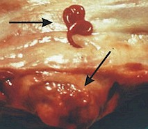
Spirocerca lupi, the esophageal worm, is a parasitic roundworm of dogs (very occasionally cats) and other carnivores (e.g. foxes, wolves, coyotes, lynxes, etc.).
It is found worldwide in tropical and subtropical regions, including the southern United States and Mediterranean Europe. Studies in Greece found that up to 20% of hunting dogs can be infected, whereas no infections were recorded in household dogs.
Spirocerca arctica, is a related species found in cold regions in northern Europe and Asia.
The disease caused by Spirocerca lupi is called spirocercosis.
Spirocerca lupi does not affect cattle, sheep or goats.
Are dogs infected with Spirocerca lupi contagious for humans?
- NO. Neither through contact, nor through the feces or the vomit. The reason is that this worm species is not a human parasite. For additional information read the chapter on the life cycle below.
You can find additional information in this site on the general biology of parasitic worms and/or roundworms.
Final location of Spirocerca lupi
Predilection site of adult Spirocerca lupi is the esophagus, occasionally the stomach and the aorta.
Anatomy of Spirocerca lupi
Spirocerca lupi is a medium-sized roundworm, about 4 to 8 cm long and have a pink to red color, often coiled. The worm's body is covered with a cuticle, which is flexible but rather tough. The worms have no external signs of segmentation. They have a tubular digestive system with two openings. They also have a nervous system but no excretory organs and no circulatory system, i.e. neither a heart nor blood vessels. Males have chitinous spicules for attaching to the female during copulation.
The eggs are elliptical, rather small (~35x14 micrometers) and contain already infective larvae when shed in the feces.
Life cycle and biology of Spirocerca lupi

Spirocerca lupi has an indirect life cycle with dogs and other carnivores as final hosts, coprophagous beetles (e.g. Geotrupes spp, Skarabeus spp) as intermediate hosts, and numerous vertebrates (chicken and other birds, frogs, lizards and other reptiles, rodents, pigs, etc.) as transport hosts.
Adult females lay eggs that are shed with the host's feces and contain infective L1 larvae. The beetles eat these eggs and the released larvae develop to L3 larvae in the host's body and encyst. Transport hosts eat the beetles, the L3 larvae are released in their stomach and they encyst again without completing development.
Dogs (very occasionally cats as well) and other carnivores become infected mainly after eating infected transport hosts. The encysted larvae are released after digestion, penetrate into the blood vessels of the stomach and migrate further to the thoracic aorta where they arrive within about three weeks. They remain there for about 2 to 3 months. Subsequently most larvae migrate to the esophagus wall (occasionally to the stomach wall), where they form nodules.
Nodules are formed as a reaction of the host's organism to the intruders. It tries to isolate the worm producing connective tissue around it. Eventually the worm is encapsulated in the nodule, which remains connected to the esophagus lumen through a hole. The larvae complete development to adults inside the nodule and start laying eggs that leave the nodule through the hole. These eggs are shed with the feces.
During their migration and inside the nodules the worms feed on host's tissues, body fluids and also blood.
The time between infection and shedding of the first eggs in the feces (prepatent period) is 5 to 6 months.
Harm caused by Spirocerca lupi infections, symptoms and diagnosis
Infections with Spirocerca lupi can be serious, even if the may not cause clinical signs. The nodules in the esophagus can hinder swallowing and cause vomiting and hypersalivation or they can impair breathing through partial obstruction of the adjacent airways. Nodules in the blood vessels can disturb the blood flow as well.
Larvae in the wall of the blood vessels can cause aneurysms, which usually heal but leave scars that impair the vascular functionality. But some aneurysms may burst and cause the sudden death of the host without previous warning.
In addition, the nodules in the esophagus or elsewhere often develop into cancer that can spread to other organs. Another possible development, even before respiratory or digestive signs, is hypertrophic osteopathy, which is characterized by thickening of the long bones and leg swelling of affected dogs.
For diagnosis, endoscopy, radiography or echography can visualize the nodules or individual worms. Fecal examination for eggs is not reliable, because egg shedding in the feces is intermittent, which can produce false negatives.
Prevention and control of Spirocerca lupi infections
In endemic regions dogs should be prevented from eating potential intermediate hosts (beetles) or transport hosts (lizards, frogs, rodents, birds including raw chicken, etc.), but this is rather difficult to achieve with hunting or working dogs. In kennels and boarding houses feces and vomits must be thoroughly eliminated as soon as possible.
Most usual dewormers for dogs are not approved for use against Spirocerca lupi. It is known that doramectin and ivermectin can be effective, but dose and treatment regime have to be determined by the veterinary doctor. However, whatever dewormer will not heal aneurysms or tumors that may have developed due to the worm infection.
There are so far no true vaccines against Spirocerca lupi. To learn more about vaccines against parasites of livestock and pets click here.
Biological control of Spirocerca lupi (i.e. using its natural enemies) is so far not feasible.
You may be interested in an article in this site on medicinal plants against external and internal parasites.
Resistance of Spirocerca lupi to anthelmintics
So far there are no reports on resistance of Spirocerca lupi to anthelmintics.
This means that if an anthelmintic fails to achieve the expected efficacy, chance is very high that either the product was unsuited for the control of Spirocerca lupi, or it was used incorrectly.
|
Ask your veterinary doctor! If available, follow more specific national or regional recommendations for Spirocerca lupi control. |